This technique has been selected for oral presentation in the
8th International Meeting on Microbial
Epidemiological Markers (IMMEM-8), to be held in Zakopane,
Poland, May 14-17, 2008:
Plenary session #1:
New Typing Strategies and Technologies: Oral presentation #6
Application of resAP-PCR
fingerprinting to strains from sequenced bacterial species
I. Martinez-Ballesteros, A. Bernal, R. San Millán, A.
Rementeria, J. Garaizar, J.
Bikandi
|
This technique was primarely developed by in silico simulation, and latter
it was applied to Salmonella strains.
The performance was excelent. The methods used are described bellow:
-
Organisms.
A total of 27 Salmonella strains (check table bellow),
grown on trypticasein soy agar at 37 ºC
overnight, were
examined. The cultures were obtained
from our collection of Salmonella
spp. strains at the Department of Immunology, Microbiology and
Parasitology,
University of the Basque Country. They belong to 13 serotypes of Salmonella enterica.
|
Serotype
|
Strain number
|
Serotype
|
Strain number
|
|
Typhimurium
|
LT2
|
Anatum
|
331
|
|
Typhimurium
|
75
|
Anatum
|
342
|
|
Typhimurium 4,5,12:i:-
|
2B-
|
California
|
327
|
|
Enteritidis
|
26
|
California
|
333
|
|
Enteritidis
|
28
|
Derby
|
343
|
|
Miami
|
261
|
Derby
|
345
|
|
Miami
|
8
|
Lexington
|
367
|
|
Arizonae
|
83
|
Lexington
|
378
|
|
Arizonae
|
20
|
Llandoff
|
334
|
|
Hadar
|
272
|
Llandoff
|
335
|
|
Hadar
|
275
|
Kentucky
|
531
|
|
Montevideo
|
328
|
Kentucky
|
534
|
|
Montevideo
|
454
|
Agona
|
263
|
|
|
|
Agona
|
337
|
- DNA extraction. Bacterial colonies were
resuspended in saline solution until a 2 McFarland concentration and
1ml of
that solution was used for the DNA extraction. The DNA extraction was
performed
according to the protocol supplied in the DNeasy Blood and Tissue kit
(Qiagen).
The bacterial DNA was recovered in 200 µl of elution buffer
supplied
with the
DNeasy Blood and Tissue kit. To this protocol it was added a DNA
concentration
phase with Sodium Acetate 3M. To determine the amount of DNA,
1 µl of
each
sample was measure with the NanoDrop ND-1000 Spectrophotometer.
-
Digestion.1
µg
of genomic DNA was digested with
10U of Hae III in 10X M Buffer
(final volume, 10 µl) for 2h at 37ºC.
- Selection
of Oligonucleotides for PCR amplification. The procedure is
described bellow.
- PCR Amplification. Each 25 µl PCR mixture included
1 µl of template
DNA (digested genomic DNA), 1U of Taq DNA polymerase (BIOLINE),
deoxynucleoside
triphosphates (200 µM each) (BIOLINE), 2.5 mM MgCl2 and
the
three oligonucleotide
primers (1 µM) in 1X PCR buffer. Amplification was
performed in a
RoboCycler 40
(Stratagene) with an amplification profile that consisted of an initial
denaturation step at 95ºC
for 2 min and then 30 cycles with denaturation at 95ºC
for 1 min,
primer
annealing at 32ºC
for 30 s, and extension at 72ºC
for 1 min. To ensure complete strand extension, the reaction mixture
was kept
at 72ºC
for 4 min after the final cycle. All the experiments included negative
controls
which were processed with the samples. The amplified products were
resolved on
2% agarose gels and the bands stained by ethidium bromide staining,
visualized
in a transilluminator. The banding patterns were compared visually.
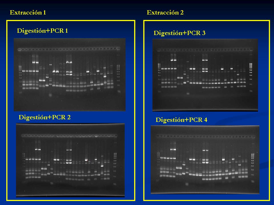
- Reproducibility of
the
technique. DNA extractions were
performed from
each strain in two separated days. Each DNA sample was used for
restriction
with HaeIII and subsequence PCR amplification and separation in agarose
gel in
two different days. In total, four gel images were obtained to search
for
reproducibility. The images are shown
bellow:
Selection of primers
- Software. The programs used for selection of
oligonucleotides were generated by our group. PHP scripting language
was used
to develop them, and they were run in an Apache/PHP environment
- Selection of
oligonucleotides. The
procedure for selection of 9 nucleotides long primers was performed in
5 steps:
- Selection of 9 bases long primers with Tm over
30 C and
no hairpin or other unwished structure formation. A total of 37,913
oligonucleotides were selected.
- Selection of primers matching the genome of
Salmonella
Typhimurium LT2 at least 200 times after its theoretical restriction
digest with HaeIII (GG'CC).
- Search for trios of primers from the selected
ones in the
previous step. Only trios yielding at least 8 theoretical bands by PCR
for Salmonella Typhimurium LT2 cleaved with HaeIII were selected. The
number of trios fulfilling the minimal bands requirement was 315, from
which 81 had an A+T content equal to 2, and in 234 was equal to 3.
- Search for PCR amplification with trios selected
in
previous step: Salmonella enterica subsp. enterica serovar Typhi
(NC_003198), Salmonella typhi Ty2 (NC_004631), and Salmonella enterica
subsp. enterica serovar Paratyphi A str. ATCC 9150 (NC_006511).
- Selection of trios of primers for wet
experiments. The
selection was visual, and no specific selection procedure was applied.
PCR amplification with the proposed technique
was applied to 27 strains of Salmonella.
- As wet lab results were
excelent, the
procedure was applied to all sequenced prokaryiotes. For some
species
the technique is not applicable (there are no oligonucleotide trios
available), but this genotyping technique may be applied to most
pathogens.
|